Image
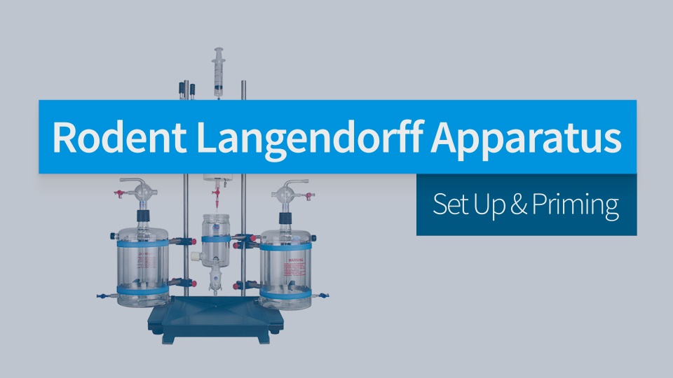
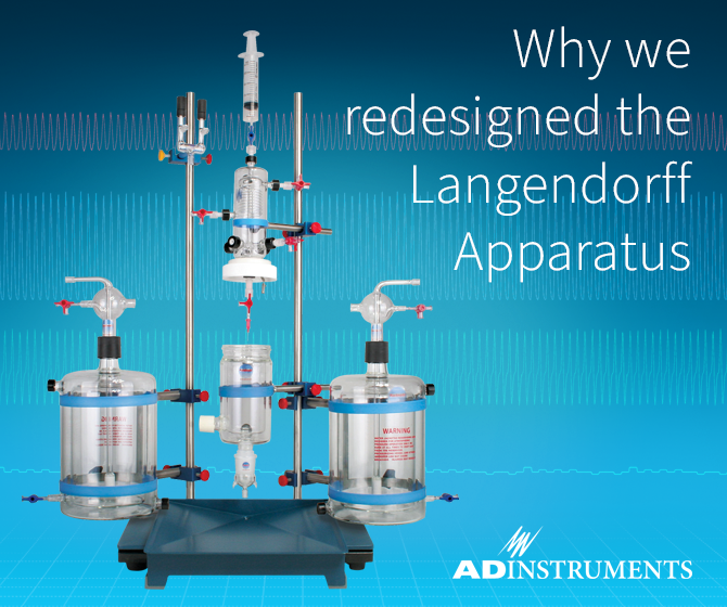
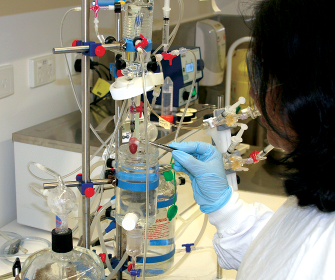
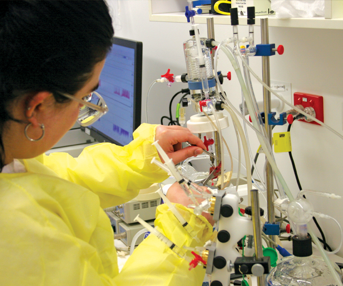
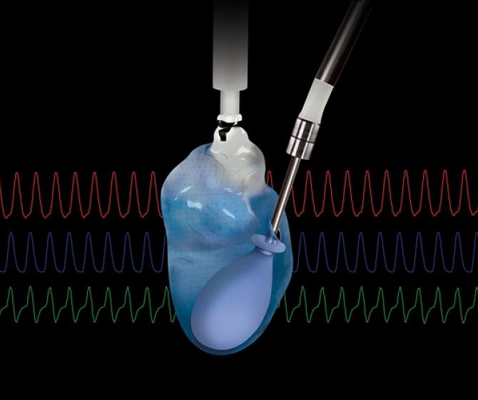
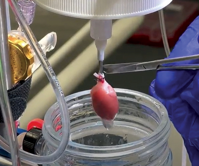
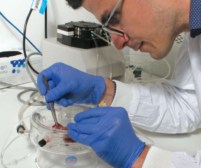
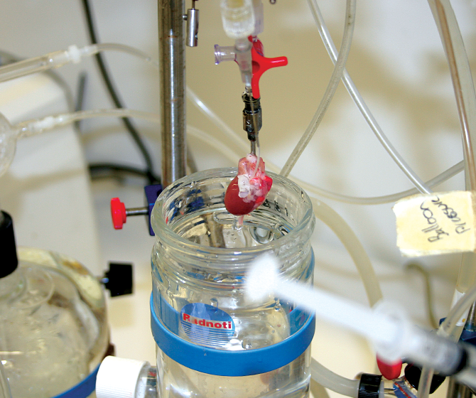
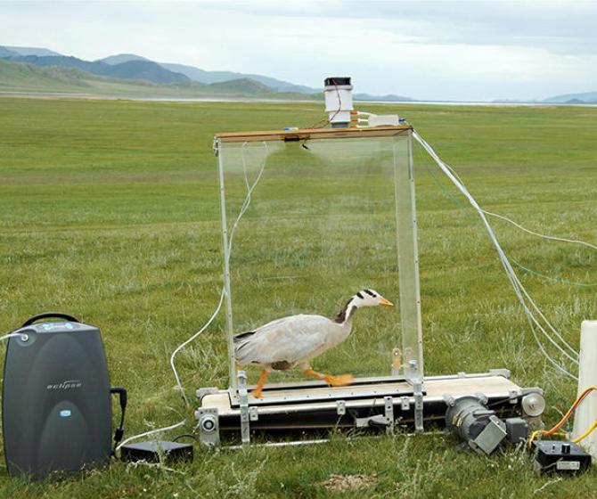
Cardiovascular research focuses on physiology of the heart and cardiovascular system. From basic biological investigation through to disease focused research, and translational research with a bench to bedside focus, cardiovascular research in animals is a diverse and multidisciplinary application.
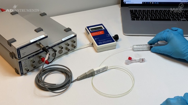
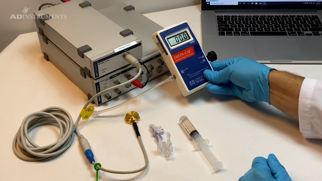
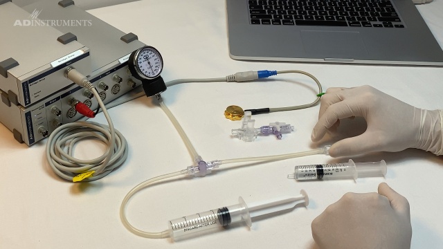
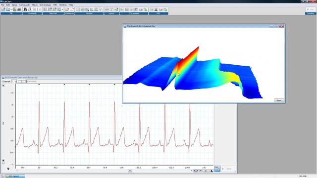
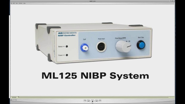
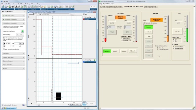
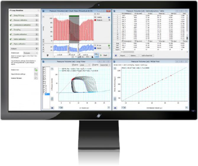
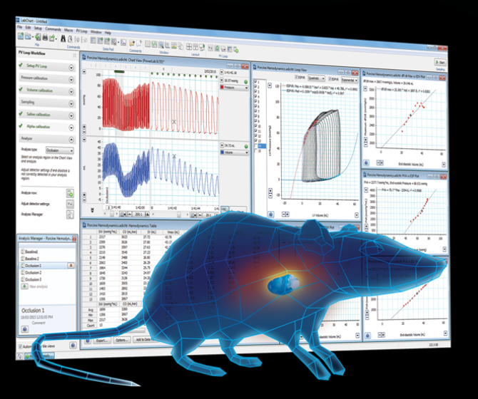
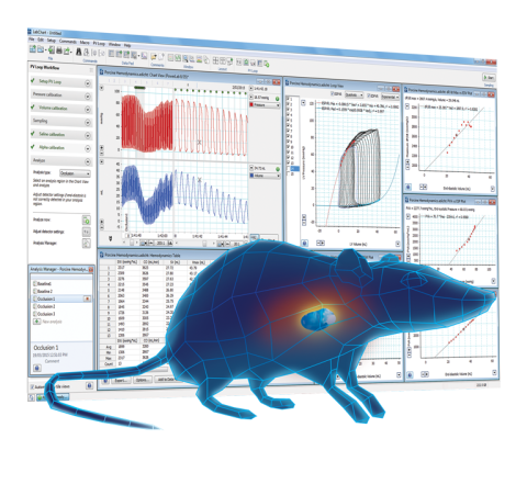
Whether your research is basic or translational, or somewhere in between, ADInstruments offers a range of solutions for accurate and sensitive cardiovascular measurements. And with the ability to integrate data streams from blood flow, NIBP, isolated heart, arterial pressure, ventricular pressure and volume, laser doppler flow, electrophysiology and more, our systems can evolve as your experiments do - ensuring quality results wherever your research takes you.