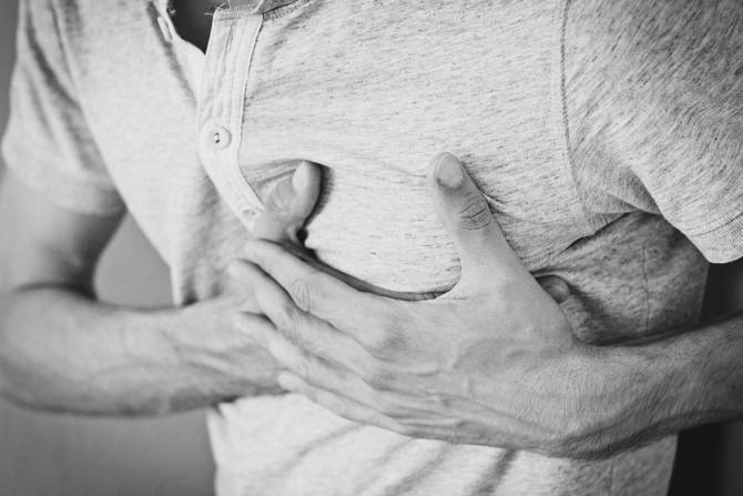
Tightness of the chest. Shortness of breath. Pain in the jaw, arm or back - am I having a heart attack!?
While many of us are familiar with the signs and symptoms - we have no clue as to what is actually happening to the heart during a heart attack aka myocardial infarction. In fact - many individuals won't regain full cardiac function after experiencing one. While current therapies focused on restoring blood flow to the heart have proved successful, the residual myocardial scarring that often remains is impairing patients from regaining full cardiac function. This leaves them at risk of heart failure... Not good.
The use of stem cells, macromolecules or small molecules has been a hot topic in this area known as cardiac regenerative therapy. However, limitations in the delivery and viability of the transplanted material to the post-myocardial infarcted (MI) heart is an ongoing problem. In order to address these issues, a collaborative team of researchers from NUI Galway, Harvard University, Massachusetts Institute of Technology, Trinity, RCSI and AMBER have developed a therapeutic epicardial device called Therepi. By combining Millar's highly sensitive, pressure-volume catheters with PowerLab data acquisition and LabChart data analysis software, the team were able to accurately report on the promising tissue regenerative effects of the Therepi system.
Development of the Therepi System
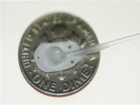
Therepi's tiny reservior.
The purpose of Therepi is to allow for sustained and repeated delivery of small molecules, macromolecules and cells directly to the damaged heart via a polymer-based reservoir that is connected to a subcutaneous port. More importantly, this implantable system allows for multiple administrations of the desired therapy to promote tissue restoration whilst simultaneously eliminating the need for multiple surgeries. The study was published last year in the internationally respected journal, Nature Biomedical Engineering. Click here to read more about the design and manufacturing details of the Therepi system.
Testing the viability of the Therepi system in a rat model
Whyte et al. tested the potential of the Therepi system in a rat model. A thoracotomy was used to insert the device, during which time the left anterior descending artery was permanently ligated to induce a myocardial infarction. A detailed description of the surgical method used is available in the supplementary information.
To test the viability of Therepi as a treatment delivery system over a 28 day study period, they looked at:
- Targeted delivery of small molecules - using the imaging substrate D-luciferin
- Localization of delivered material i.e cells, paracrine factors and other macromolecules
- The ability of macromolecules to penetrate the myocardial tissue to the target area
- The effect of repeated cell delivery through the port on the resident cell population
- Whether cardiac function improved with Therepi based cell-refilling i.e. repeated dosing
Five different treatment groups were used to determine the effects of the Therepi system on cardiac function over the 28 day study; (1) sham (MI only), (2) a single injection of cells, (3) Therepi without any cells, (4) Therepi with cells and (5) Therepi with cells and refills.
Using ventricular pressure-volume (PV) loop analysis to measure changes in cardiac function
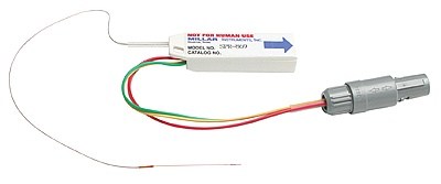
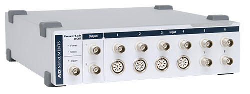
To measure the efficacy of the delivery system on improving cardiac function, they performed functional haemodynamic measurements using an echo ultrasound and a PV catheter (Millar Mikro-Tip) for each of the test groups. Ventricular pressure and volume (PV) were measured using a conductance catheter (SPR-838) that had been inserted into the left ventricle via the apical stick method. From here, PV data was obtained and analysed using the Powerlab 8/35 and LabChart Pro 8 software with the PV module. PV-loops generated from the different treatment groups could then be compared, and data such as the ejection fraction and stroke work were plotted as a means of assesing changes in cardiac function.
This type of analysis highlighted the effect that Therepi + cells + refills had on increasing the ejection fraction and stroke work of the heart, indicating a positive healing effect from the treatment.
Key findings
This comprehensive study not only included the design of the Therepi system, but also a pre-clinical trial in a rat model, highlighting the potential for this system to be used in the clinical setting. Overall, the results of this study showed that Therepi is able to:
- Effectively deliver small molecules to the diseased area that can either have a functional effect, or be used for in vivo imaging, to quantify cell numbers on the heart
- Enable localized delivery of macromolecules that can penetrate the fibrous heart tissue and target the diseased area in three-dimensions.
- Provide a mechanism for non-invasive replenishment of cells in vivo, improving cardaic regeneration by increasing cell number at the target site by up to 10-fold
While the best results were seen in the test group that had the Therepi system plus multiple doses over the 28-day period, slight improvements in cardiac function were seen in the Therepi only control group (2). Interestingly, the presence of the Therepi system alone was thought to aid in the healing of the heart by providing either; mechanical reinforcement for the remodelling of the heart tissue or stimulate an altered healing response due to the presence of a foreign body.
This innovative new piece of biotechnology designed by Whyte et al. provides a viable mechanism for the clinical translation of cardiac regenerative therapy, as well as the potential to be used as drug delivery system for the treatment of other diseases and health issues.
So what's next?
While this is a promising new piece of technology in both research and clinical fields, Whyte mentioned that future work will focus on developing a biodegradable device, so that no foreign material remains on the heart post-treatment. There was also mention of a permanent device that would allow for acute therapy replenishments initially as well as encompassing the long-term passive reinforcement effects mentioned above.