Kaha Sciences biopotential telemeters provide an excellent tool for collecting high quality, low noise ECG signals for long durations in conscious animals. To obtain high quality recordings correct placement of electrodes is important. The following is our recommendation for attaching the leads to obtain a modified lead II ECG signal.
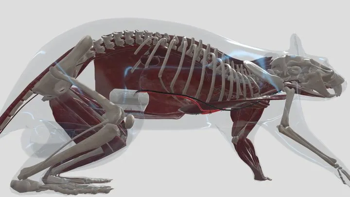
As the animals will be conscious and moving around, the reduction and elimination of interfering electrical signals is important. In general, low noise ECG signals can be obtained if the electrodes are located close to the heart — along the electrical axis of the heart with the electrode tip embedded in the muscle layer rather than “floating” under the skin. For most animals, the frequently used lead II configuration provides large amplitude deflections that are easily recognized both visually and are suitable for analysis by ECG analysis software.
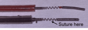
Telemeters are supplied with bipolar electrodes that are well suited to ECG data collection. The electrode leads are manufactured from high quality stainless steel (316 grade) using a manufacturing process similar to that used to construct pacemaker leads for use in humans. This gives the leads high tensile strength while still having a high degree of flexibility. The leads are inside medical grade silicone tubing with the last 5 mm of electrode lead exposed. Should the electrode tip become damaged it is possible to trim back the silicone tubing using a scalpel to expose a “fresh” section of electrode tip.
Electrodes are supplied connected together down their entire length of the silicone. While separation of the ends is required to allow placement, we recommend that the electrodes be kept together and run alongside one another for as far as practical. A free length of no more than 3-4 cm should be needed for each electrode.
It is not recommended that sutures be placed around the leads other than at the point of contact with the tissue as this may provide a stress point on the leads.
Electrode Placement
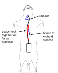
For signals of the best quality, we recommend the use of the method developed by Sgoifo et al. (Physiology & Behaviour vol 60, pp1397-1401, 1996). This approach to electrode lead placement seeks to reduce artefact associated with breathing and movement. After placing the telemeter within the abdominal cavity one lead is fixed to the dorsal surface of the xyphoid process. The other electrode is subcutaneously tunneled through to the upper insertion of the sternohyoid muscle. The electrode wire is formed into a U shape, pushed under the muscle and along the trachea into the anterior mediastinum, to get the recording lead as close to the right atrium as possible.
Key points:
- Ensure electrodes are securely fastened using sutures on both the silicone tubing and the exposed electrode wire.
- The exposed coiled electrode wire should be stretched at the suturing point and the suture tied between two coil loops. This will ensure that the wire cannot pull through the suture.
- The two electrode wires should be kept close together for as far as practical.
- Only suture the electrode wires at the points of contact.
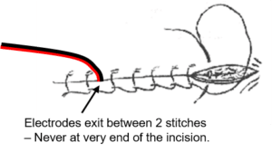
- The electrodes should always exit the abdominal cavity between two stitches and never at the very end where they may cause separation of the abdominal muscles over time.
Videos contain content hosted by Millar prior to the formation of Kaha Sciences. Kaha Sciences is an ADInstruments brand. Any contact details provided in videos are now out of date - For all product information or support, please contact us.