×

Intracellular Recordings

Intracellular Recordings

Intracellular recording is an electrophysiology technique that inserts a glass microelectrode into a single cell (usually a neuron) to precisely measure its electrical activity (voltages across or currents passing through the cellular membranes). It can allow the recordings of slower graded voltages (receptor potentials or synaptic potentials), which would be vital in studies involving neural signalling.

Ask a Question Book a Demo

Intracellular Recordings

Signal Overview

There are several variations of intracellular techniques, which can be grouped by the type of glass microelectrode used.  The first group uses a microelectrode with a relatively small tip (fine or sharp), while the second uses a relatively large tip (blunt or patch). Each technique uses Ohm’s Law to focus on various aspects of the electrical activity.

Sharp Electrode
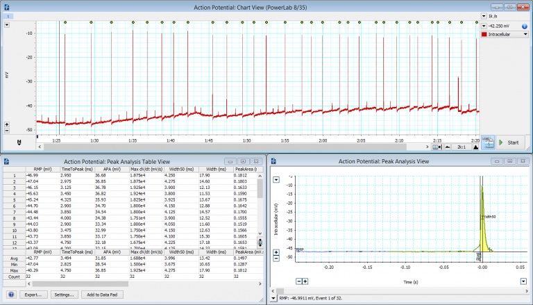
Membrane Potential/Voltage (Current Clamping): This technique measures the membrane potential by injecting a constant electrical current through the recording electrode, which is usually done to probe the passive or active properties of the cell membrane. The amplifier involved is designed to compensate for any resistive artifacts by the microelectrode. Current injection can also be used to introduce charged substances into the cell (iontophoresis), which can be used in cell marking techniques.

Membrane Current (Voltage Clamping): Classical voltage clamping involves the use of two electrodes (Two Electrode Voltage Clamping or TEVC), whereby one electrode records intracellular voltage and the other electrode injects current. The voltage-clamp amplifier forms a feedback circuit between the recorded voltage and the injected current. This then allows changing the cell membrane voltage to various held values, while recording the associated injected current required to realize the corresponding ‘clamped’ voltage. TEVC is most commonly used on large cells such as oocytes, and thus is sometimes referred to as oocyte clamping. Another voltage clamping technique is Single Electrode Voltage Clamping (SEVC), or sometimes known as switched clamp, uses a time-sharing technique that alternates between recording voltage and injecting current in a single intracellular electrode. It uses high frequency digital switching circuits, where the recording time is a short and fast enough that the cell remains ‘clamped’.

Patch Clamping
Unlike conventional sharp electrode techniques that involves impaling a cell, the patch microelectrode is placed next to a cell, and gentle suction is applied to draw a piece of the cell membrane (known as the 'patch') into the microelectrode tip, creating a high resistance (gigaohm) seal with the cell membrane. Variations on the suction can provide a variety of patching techniques including cell-attached, whole cell (similar to sharp electrode), outside-out patch and inside-out patch. Depending on the patching techniques, currents passing through individual ion channels, or through the whole cell can be recorded, or pharmacologically analysing the patch’s membrane properties.

Signal
LabChart

Enabling Discovery

LabChart

All your analysis in one place

LabChart software is designed specifically for life science data and provides up to 32 channels for data display and analysis options that are powerful and easy to use. With auto-recognition of ADI and LabChart Compatible hardware, multi-window views, one touch recording, simultaneous recording from multiple devices, specialized preconfigured settings, easy sharing options and an interface that can be customized to show only the features you want to use.

Noise Elimination

This is a common issue in electrophysiology as you are dealing with very small signals as well as high impedance and capacitance of electrodes. All of ADInstruments hardware is optimized with high-quality noise specifications, but it is always good to have a few other tricks up your sleeve to help in your setup. 

  1. Ensure all electronic equipment used in recording setup is plugged into the same wall socket by using a fused multibox.
  2. Ensure all shielding is connected to one grounding point or grounding bar. This will eliminate the risk of a grounding loop when different grounds are at slightly different potentials causing currents to flow in the shielding introducing noise.
  3. To identify the source of noise, strip back to the bare essentials, like the amplifier and acquisition and then systematically switch on additional equipment.
  4. Always try to eliminate the source of noise first, but if this isn’t possible then mask with shielding.
  5. Try and keep lead wires as short as possible and not looped. If they are long, try braiding them to prevent magnetically induced currents.
  6. Even fluid filled tubing for perfusions can act as antennas. If this is the case, shield with tin foil and connect to the metal grounding bar.
×

Features and Add-Ons

Additional acquisition and analysis options to support your Intracellular Recordings analysis:

LabChart Lightning
LabChart Lightning
Play video

Data acquisition and analysis re-imagined.

Make unique discoveries with unlimited freedom and flexibility.

Try LabChart Lightning now for free

This site is protected by reCAPTCHA and the Google Privacy Policy and Terms of Service apply.

Oops, something went wrong...

Thanks, we've sent you an email with instructions on how to start your trial.

Request a demo

Thank you for your interest in Intracellular Recordings. Please complete and submit the form below and an ADInstruments representative will aim to get in touch within one working day. If you’d like to speak to someone immediately, office contact information is available from the contact page.

×

Ask a question

Thank you for your interest in Intracellular Recordings. Please complete and submit the form below and an ADInstruments representative will aim to get in touch within one working day. If you’d like to speak to someone immediately, office contact information is available from the contact page.

×