×

Electrical Stimulation

Electrical Stimulation

Electrical stimulation has long been used in research to either observe propagation or activation properties, mapping and threshold studies in both human and animal subjects. By varying the amplitude, duration and frequency of stimulation, it has been used both in vivo and in vitro, to study various tissues and organs including muscle, brain, heart, and the nervous system.

Ask a Question Book a Demo

Electrical Stimulation

Signal Overview

Electrical stimulation has been used in various in vivo and in vitro physiological studies and applications, which include studies on muscle contraction and relaxation, muscle activation or thresholds, nerve conduction or propagation, and neural networks. Variations of electrical stimulation amplitude, duration, frequency and patterns have been used on a range of tissues and organs from various species have been studied including muscle, brain, heart, and the nervous system.

In isolated tissue-organ bath studies, electrical stimulation may be directly applied or indirectly applied via field stimulation to induce isometric or isotonic contractions in skeletal, smooth and cardiac muscle. Sufficient current passing through the tissue sample needs to be ensured, which depends on the tissue sample resistance. Skeletal muscle contraction studies may also include summation and/or tetanisation protocols. In whole intact heart experiments, electrical stimulation can be used for pacing.

Noninvasively, electrical stimulation can also be used to examine the resultant force and recruitment of skeletal muscles and its application is also known as electrical muscle stimulation (EMS).

Neuromuscular relationships can also be studied, which allows the study of nerve conduction in terms of timing and velocity. Furthermore, invasive studies of action potential propagation with the use of electrical stimulation can also be performed, which can also allow identifying and mapping of neural networks.
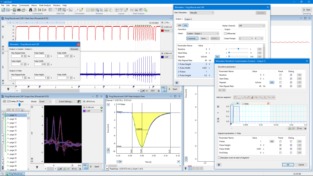
Signal
LabChart

Enabling Discovery

LabChart

All your analysis in one place

LabChart software is designed specifically for life science data and provides up to 32 channels for data display and analysis options that are powerful and easy to use. With auto-recognition of ADI and LabChart Compatible hardware, multi-window views, one touch recording, simultaneous recording from multiple devices, specialized preconfigured settings, easy sharing options and an interface that can be customized to show only the features you want to use.

Isolated tissue applications

In isolated tissue applications, current driven simulators are often a better choice, as variation in the path through the tissue may produce too much variability when simple voltage control is use. In addition,  where multiple preparations or samples are to be stimulated using a single stimulator, care must be taken to ensure that the current is not split unevenly between different baths or preparation.

×

Features and Add-Ons

Additional acquisition and analysis options to support your Electrical Stimulation analysis:

LabChart Lightning
LabChart Lightning
Play video

Data acquisition and analysis re-imagined.

Make unique discoveries with unlimited freedom and flexibility.

Try LabChart Lightning now for free

This site is protected by reCAPTCHA and the Google Privacy Policy and Terms of Service apply.

Oops, something went wrong...

Thanks, we've sent you an email with instructions on how to start your trial.

Request a demo

Thank you for your interest in Electrical Stimulation. Please complete and submit the form below and an ADInstruments representative will aim to get in touch within one working day. If you’d like to speak to someone immediately, office contact information is available from the contact page.

×

Ask a question

Thank you for your interest in Electrical Stimulation. Please complete and submit the form below and an ADInstruments representative will aim to get in touch within one working day. If you’d like to speak to someone immediately, office contact information is available from the contact page.

×