In extracellular electrophysiological recordings, an electrode is not inserted into a single cell, instead the electrode(s) are place in the extracellular fluid, near the cell of interest. The type of recording will depend on the type and size of the electrode, and proximity to the signal origin.
Single/Multiple Unit Recordings
In these types of extracellular recordings of action potentials, instruments sample the electrical activity of a single neuron or several neurons at once. Recorded activity is referred to as unit as you cannot be sure if spikes are in fact action potentials. Recordings can be made either in vivo or in vitro. To record these types of signals, a glass or metal electrode with relatively high impedance is used alongside a headstage with high input impedance. If the electrode is very small (+ 1um tip), the activity of a single adjacent neuro can be recorded. If a slightly larger tip is used, it will record the activity of several neurons adjacent to the electrode, and spike sorting needs to be employed to identify particular cells.
Epithelial Voltage Clamping (and Ussing Chambers)
In order to measure the transport of electrolytes, non-electrolytes and H2O across an epithelial membrane, an Ussing chamber setup is required. The chamber isolates each side of the membrane so that they face a separate chamber half so that chemical and electrical adjustments can be made to each side separately. With a Ussing setup (usually comprising a chamber, a perfusion system, an amplifier, and an acquisitions and analysis system), measurements can be taken from native tissue including stomach, large and small intestines, gall and urinary bladder, skin and trachea, as well as from tissue derived cell monolayers from various sources including renal tubes, pancreas, and salivary and sweat glands.
Whole Nerve Recordings
Many researchers are interested in recording the activity of peripheral nerves, which are essentially bundles of axons. This can be done with wired methodology where an electrode (usually silver wire) is placed in contact with the nerve in question. Another methodology is through the use of telemetry products where sympathetic nerve activity can be recorded.
Field Potentials
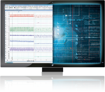
Field potentials are probably the most common extracellular signals being recorded and include ECG, EMG and EEG. With an electrode with an even bigger sampling field, the activity of individual neurons can no longer be distinguished, but rather a field potential generated by the activity of many cells. Extracellular field potential recording can either be carried out invasively or non-invasively on the skin.