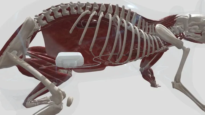
With a high fidelity solid-state pressure sensor located at the tip of the catheter, the Kaha pressure telemeters are ideal for accurately recording left ventricular pressure in rats. The surgical methods have been broken up into short sections below with the key points for each video highlighted in the text for each section.

Full details of the methods described here can be found in the publication by Stehlin et al 2013.
A recording of a webinar presenting this methods and some data examples can be found here.
Anesthesia for LVP implantation
Providing an adequate level of anesthesia is one of the critical aspects to chronic implantation of monitoring devices. The wrong anaesthetic will slow recovery or the animal may die during surgery.
The level of anesthesia should be assessed prior to and throughout surgery by monitoring the withdrawal reflex in response to a pinch of the paw pad throughout the surgery. A pulse oximeter sensor placed on the paw of the animal to measure heart rate and oxygen saturation can also be useful.
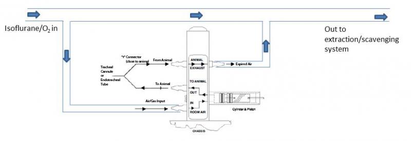
Isoflurane and other gaseous anaesthetics provide an easy and effective way of inducing and maintaining anesthesia.
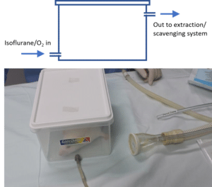
Induction: To induce anesthesia the animal can be placed inside an induction chamber or box. This box can be as simple as a plastic container with an inlet for the anaesthetic gas and an outlet to the exhaust.
Ventilation: Once induced, the animal will need to be intubated and anesthesia maintained using a ventilator. An extraction or scavenging system must be used with any gaseous anaesthetics.
Abdominal incision for LVP measurement
- The diaphragm should be accessed via a midline incision approximately 4cm long starting at the xyphoid process.
- A piece of damp gauze is placed on top of the liver to stop it from being damaged or drying out.
Retractors can then be used to hold the incision open and the liver down to give a good view of the diaphragm. - In the video below, a pressure catheter has already been inserted into the abdominal aorta and now a second catheter will be placed in the left ventricle by extending the incision up the midline to the xyphoid.
Inserting and securing the catheter in the heart
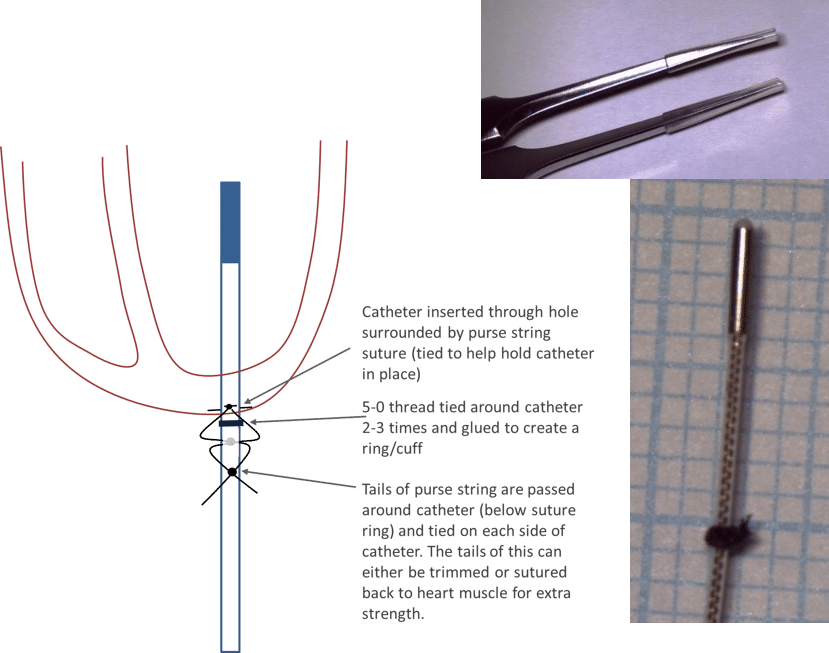
For measurement of LVP, the recommended method for securing the catheter in place is to use sutures (see figure below) rather than tissue adhesive (which may release after a period of time). A cuff/ring of suture is used to help secure the catheter the correct distance into the heart. For rats 250-450g inserting the very tip of the catheter ~10 mm into the heart (via the apex) has been found to give the most stable LVP signals.
To prevent damage, the catheter should be handled using fine forceps with the tips padded with silicone tubing.
In the video below, the apex of the heart has already been exposed through an incision in the diaphragm. The pressure catheter is now inserted into the left ventricle through a hole in the apex and secured using the method described above.
Closing the diaphragm
Once the catheter is in place, the diaphragm must be closed and the air removed to restore the negative pressure in the chest cavity.
In the video below, individual sutures are used to close the diaphragm around the catheter. As the final suture is tightened the lungs are inflated and the air is removed using a syringe and plastic canula.
Note: To allow the heart to lie in its natural position, make sure the catheter is towards the rat’s left hand side when the diaphragm is sutured closed but make sure it exits between two sutures and not at the very end of the incision.
Videos contain content hosted by Millar prior to the formation of Kaha Sciences. Kaha Sciences is an ADInstruments brand. Any contact details provided in videos are now out of date - For all product information or support, please contact us.