


Homeostasis, the stability of our normal bodily functions, is not static; it is a constant balancing act.
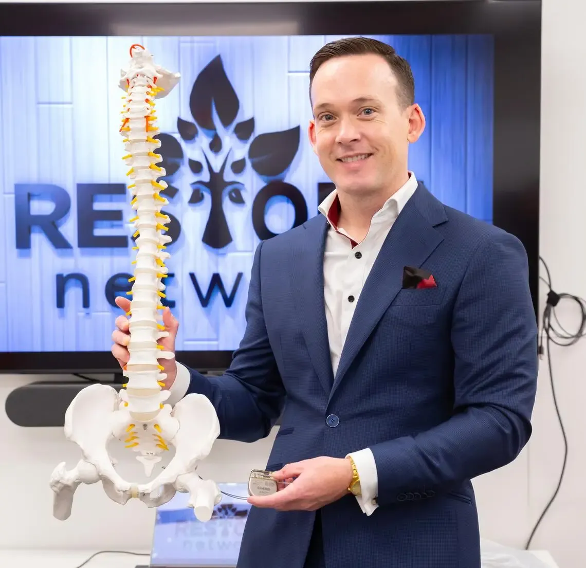
Associate Professor Aaron Phillips
Photo by Riley Brandt, University of Calgary
Information flows endlessly back and forth between the brain and the rest of the body via the spine and cranial nerves, tracking fluctuations and adjusting autonomic systems. Spinal cord injuries alter or sever that feedback mechanism and can send the process of homeostasis dangerously off course.
Autonomic dysreflexia is the adjustment of blood pressure in response to a neural circuit that didn’t exist before the spinal cord injury. What would previously have been a sensation now triggers an extreme hypertensive episode, leading to symptoms as mild as sweating or as severe as stroke, seizure, or cardiac arrest. For those living with a spinal cord injury, this life-threatening event could be brought on at any time.

Pressure, damage, or infection in the lower abdomen can trigger an autonomic dysreflexia event.
In partnered articles published in Nature and Nature Medicine, Professor Aaron Phillips and his team describe the neuronal architecture that underlies autonomic dysreflexia and the electrical stimulation protocol they have developed to treat it. These articles come just four years after the team’s work on the neuroprosthetic baroreflex, an epidural stimulator which reliably stabilised unstable blood pressure after spinal cord injury. These new publications build on the groundwork laid by the neuroprosthetic baroreflex, examining the ‘hemodynamic hotspots’ it targeted even more precisely, and using that information to fine-tune the team’s use of epidural electrical stimulation as a treatment.
Related: Hemodynamic Instability: Solving the invisible problem
These two articles are densely packed with highly skilled and precise experiments, and detail a scientific journey that covers every step the team took from developing an animal model of autonomic dysreflexia through to testing a stimulator implanted in human participants who live with the condition. In this work, Aaron and his team achieve a feat that feels near impossible in most of science: They build a complete view of the problem, and then they fix it. In this blog, we will be covering the first of the two articles, but we highly recommend that you read them both.
Nature: A neuronal architecture underlying autonomic dysreflexia
Nature Medicine: An implantable system to restore hemodynamic stability after spinal cord injury
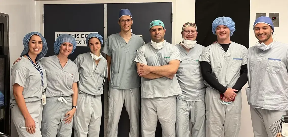
From left: Robyn Cameron, Kelly Larkin-Kaiser, Catherine Veilleux, Robin Demesmaeker, Fady Girgis, Kyle Rogan, Aaron Phillips and Nico Hankov.
A model of autonomic dysreflexia
The team began by developing a reliable clinical model of autonomic dysreflexia. In humans, the condition occurs after spinal cord injury. It tends to be triggered by persistent pressure or damage in the lower abdomen (e.g. a distended bladder or bowel, pressure sores, or infection).
A mouse model was developed with a transection of the spinal cord at T4, and mechanical distension of the colorectal area. The autonomic dysreflexia began around 2 weeks after the spinal cord injury, and peaked after 4 weeks. The delay in development suggested that the cause might be related to spinal nerve reorganization after the injury.
Identifying the neurons responsible
With an animal model, the team could reliably track changes in the spinal cord caused by autonomic dysreflexia. They cleared, labelled, imaged, and measured the amount of Fos present throughout the spine; areas with more Fos had more gene expression occurring in response to autonomic dysreflexia and were, therefore, most likely the culprits of any neuronal reorganization. There were two hotspots: segments L5-S1, and segments T11-T13.

Spinal segments L5-S1 and T11-T13 had the highest quantity of gene expression marker Fos after an autonomic dysreflexia event.
By identifying the neurotransmitters these nerves express, their responses to different molecules, using single-nucleus RNA sequencing to categorise them into various subcategories, and then running that information through a machine learning method that they developed called “Augur”, the team were able to isolate the neurons most responsive during autonomic dysreflexia.
Before the spinal cord injury, the neurons within the lumbosacral hotspot were associated with reaching and walking behaviours, and had no association with blood pressure at all. After the injury, these neurons had long projections to the thoracic hotspot, which was previously identified as a mediator of hemodynamic activity. It appeared as though these nerves, previously used for reaching and walking, had been reorganized and were now stimulating the hemodynamic hotspot. Importantly, stimulating the L5-S1 segments led to worsening autonomic dysreflexia over time.

The full spinal pathway of Autonomic Dysreflexia: a nociceptive output leaves the area of colorectal distension, travels to the lumbosacral hotspot, travels via long-projecting Vsx2 neurons to the hemodynamic hotspot, and then impacts hemodynamics through sympathetic preganglionic ChAT neurons.
Developing a treatment
The team repeated their tracing of neuronal subpopulations, this time focusing on neurons activated after stimulation of the hemodynamic hotspots that they had previously demonstrated could restore hemodynamic stability. They found that epidural electrical stimulation of the hemodynamic hotspots recruited proprioceptive neurons to activate the same ChAT neurons being activated during autonomic dysreflexia. This meant that the epidural electrical stimulation of that area could work to override the autonomic dysreflexia, rehabilitating the circuitry itself and eliminating the symptoms of the disorder.
This could be unpredictable and dangerous. Long-term monitoring was necessary to prove that symptoms could be properly managed, and that this treatment wouldn’t lead to further neural restructuring that would bring back or worsen the autonomic dysreflexia.
Prolonged Safety Testing
To perform safety testing, the team needed a model of the disease, the treatment, and the ability to do long-term continuous blood pressure monitoring. Using their existing rat model of spinal cord injury, the team induced autonomic dysreflexia and then began treatment using epidural electrical stimulation. For six weeks, the rats' blood pressure was continuously monitored using Kaha SNA and Pressure Telemeters. For a disorder that is both progressive in development, and can spontaneously intensify, it was particularly important that this monitoring was continuous over an extended period.
Over the course of those six weeks, the team saw progressive improvements in blood pressure management until the system was stabilised and autonomic dysreflexia was eliminated. The neurorehabilitation was successful.

An epidural electrical stimulator (EES) was implanted in the rat, stimulating the hemodynamic hotspot from T11-T13, while a Kaha telemeter provided continuous blood pressure monitoring over six weeks.
Related: Our free guide: Kaha Telemetry for Neurophysiology
Human Translation
Autonomic dysreflexia affects roughly 80% of people with tetraplegia. Of those being treated for the dysfunction, more than 90% still experience symptoms. Those symptoms aren’t always life-threatening, but they do impact quality of life; anxiety, headaches, heart palpitations, and the constant threat of a more serious side effect down the line. It is not always easy to make the jump from lab to clinic. There has to be a clear benefit to the participant and, in this case, that benefit is obvious.
In their Nature Medicine article, Aaron and his team discuss the process of testing their neurorehabilitation procedure on human participants in Canada, Switzerland, and the Netherlands, fine-tuning the process, and moving toward clinical trials in partnership with ONWARD Medical.
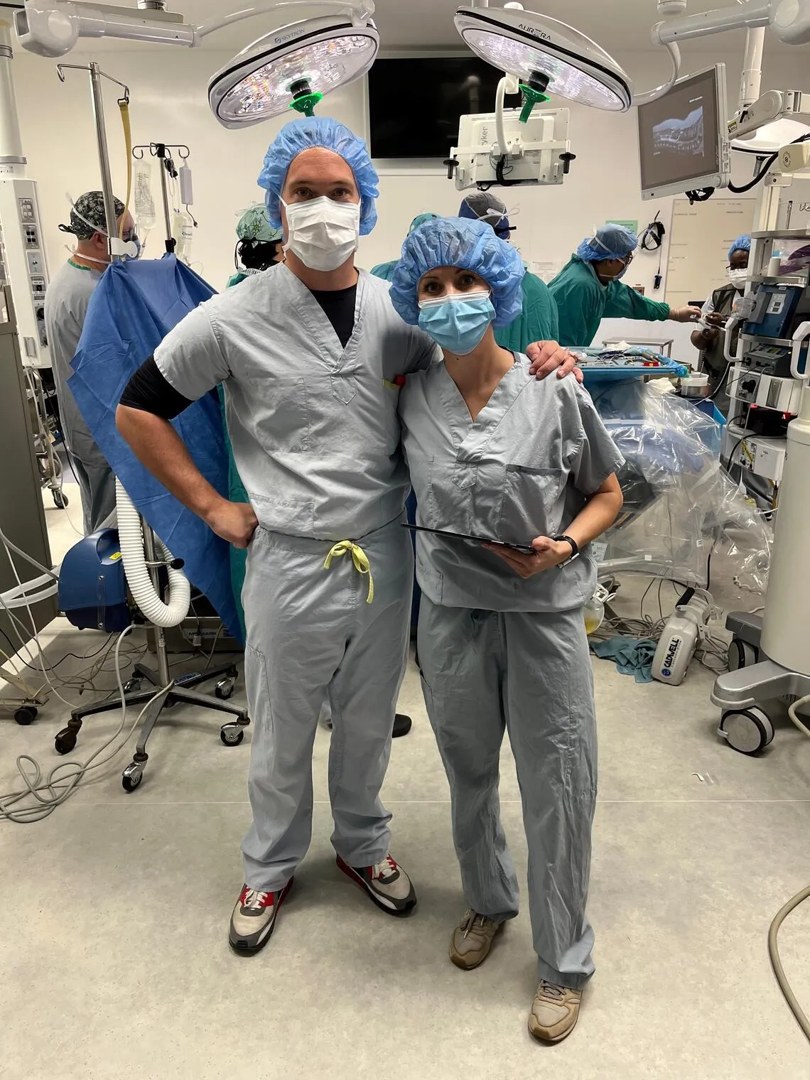
Principal Investigator Aaron Phillips, left, and Co-Investigator Kelly Larkin-Kaiser, during the first HEMO Trial Implant Surgery, August 2022.
Read More
A neuronal architecture underlying autonomic dysreflexia | Nature
An implantable system to restore hemodynamic stability after spinal cord injury | Nature Medicine
Hemodynamic Instability: Solving the invisible problem
Deep Dive: The science behind the Neuroprosthetic Baroreflex

Aaron Phillips, Ph.D.
Dr. Aaron Phillips is an Associate Professor at the University of Calgary. Driven by his passion for the complex interactions between the nervous and cardiovascular systems, including anomalous interactions contributing to clinical disorders, Aaron's research focuses on developing novel therapeutics for people with neurological health issues.