
Are you interested in measuring tissue oxygen concentrations continuously, over long periods of time? Find out how tissue oxygen can be measured in situ and why tissue oxygen telemetry could be beneficial to your research...
Overview:
1. Why is tissue oxygen an important measure?
2. Techniques for measuring tissue oxygen concentration
Why is Tissue Oxygen an important measure?
Tissue oxygen represents the balance between oxygen supply from the blood and the demand for oxygen from cells within the extracellular fluid (ECF).
Different tissues have different oxygen requirements, based on their function and needs. Even within a specific tissue, oxygen concentrations can vary based on the level of cellular activity or proximity to the capillary bed. For example, within the brain, measurements as low as 1.5 μM in can be observed in the pons and fornix, and up to 90 μM in the pia.
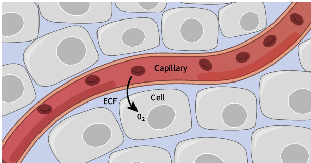
Therefore, localized measurement of tissue oxygen concentration can provide information on metabolic activity and blood flow in specific areas of the body.
Techniques for measuring tissue oxygen concentration
There are several methods used for measuring tissue oxygen concentration in-vivo. These include non-invasive techniques, such as functional imaging (fMRI and PET), and near-infrared spectroscopy.
While these imaging techniques offer a large field of view, they all require some level of animal restraint/anesthesia that can limit the types of studies performed.
Newer invasive techniques, such as implantable telemetry devices, however, can provide continuous, long-term measurements of tissue oxygen without the need for animal tethering or restraint.
Related: Introduction to Measurement of Tissue Oxygen in Rats Via Telemetry (webinar) »
How do tissue oxygen telemeters work?
Tissue oxygen telemeters work on the principle of using electrochemical sensors to measure oxygen concentration, by reducing oxygen on the surface of an electrode and measuring the resultant current.
In practice, the telemeter sensor is implanted into the desired tissue, where oxygen from the surrounding extracellular fluid is reduced, producing a current that is proportional to the concentration of oxygen within the tissue.
Where the magic happens - The carbon paste electrode (CPE)
A key component in the measurement of tissue O2 is having an electrode specifically sensitive to oxygen to ensure that other substances in the tissue are not being reduced.
The Kaha Sciences’ tissue oxygen telemeter (TR57Y) incorporates carbon paste electrodes (CPEs) within the sensor tip, to give highly sensitive measurements of tissue oxygen at the site of implantation.
Carbon paste electrodes were initially developed in the 1950’s, offering benefits over solid carbon and metallic electrodes through background noise reduction, improved sensitivity, and long-term signal stability (Bolger et al., 2010.)
Benefits of Tissue Oxygen Telemetry in your research
High quality research goes hand-in-hand with high quality equipment. You need hardware that you can trust will give you consistent and accurate results. Every. Single. Time.
These principles are at the core of Kaha Sciences’ Tissue Oxygen Telemeter design, which is why they are trusted within the scientific community as an accurate and reliable technique for measuring tissue oxygen concentration continuously, over long periods of time.
Still not convinced? Below are 4 key benefits that tissue oxygen telemetry can have for your research over other available methods...
1 - Animals can move freely and behave normally
One of the key benefits of telemetry is that it allows you to work with conscious, freely moving animals. Alternative methods involving tethers can limit animal movement, and cause behavioral changes due to the additional stress placed on the animal.
Choosing to use a fully implantable telemetry system provides an opportunity to use new experimental methods as well as improving the quality of recorded data compared to tethered applications.

2 - Enables chronic, continuous tissue oxygen measurements not possible with other techniques
Once surgically implanted, tissue oxygen telemeters can remain within the animal providing continuous O2 measurements over the course of your experiment - whether that be days, weeks, or months.
Given that some forms of dementia, stroke and cardiovascular diseases are associated with alterations in blood flow, the ability to measure oxygen concentration chronically is crucial when trying to understand the mechanisms underpinning these diseases in animal models.

3 - Back-up battery facilitates advanced study options
Kaha Sciences tissue oxygen telemeters have built-in backup batteries that allow the telemeters to continue transmitting high-fidelity data (2 kHz) for up to 4-6 hours if the animal is away from the SmartPad charging device.
The ability to continuously measure oxygen outside of the animal’s cage, such as in a water maze for behavioral studies, is a key advantage of the tissue oxygen telemeter over other available technologies.

4 -Saves you money!
Once your experiment is complete, the tissue oxygen telemeters can be surgically removed and reused for future studies, saving you money. New CPEs can be adhered to lead wires between implantations, to ensure you are recording high quality signals during every experiment.

Related: Benefits of Using Telemetry in Research »
For more information about Kaha Sciences' telemeters and how they might be useful for your particular research, please get in touch with your nearest ADInstruments Support Representative.
Additional Resources:
Solid-state sensors at the catheter tip - why settle for less? »
More about Kaha Sciences Small Animal Telemetry »
Five Unique Neuroscience Applications Using Telemetry »