Dr. Conor Underwood walks through his experience optimizing the surgical implantations of Optogenetics Biopotential Telemeter. This video is part three of our three-part interview with Dr. Underwood about his experiences.
For a complete walk-through of Dr. Underwood’s surgical process, watch our 1.5 hour Surgical Instructional video here:
Surgical Instruction: Placement of Optogenetics Telemeter within the Rat Brain »
Surgical Complexity
In Dr. Underwood’s opinion, the most difficult part of the surgical procedure is its length. Implanting the telemeter can take up to five hours. “It's really critical when you're doing a long surgery to keep everything clean. And this is especially the case if you're implanting something in an animal that's going to be in that animal for months.”
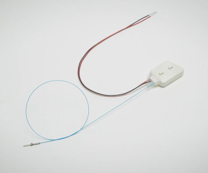
The surgical procedure involves implanting the telemeter in the abdomen of the animal, then feeding the biopotential leads and fiber optic cable a fed through a subcutaneous tunnel to the back of the head. “We are recording EEG over the motor cortex. So we make small holes above the motor cortex where we position a screw to attach the positive biopotential lead, and then another screw over the cerebellum to attach the negative biopotential lead.” After the biopotential leads are attached, the fiber optic cable is inserted into the thalamus through the skull.
Optimizing the Surgical Procedure
Changing the anesthetic was one of the most critical improvements to the surgical procedure, allowing for the animal to be properly anesthetized for the duration of the surgery. “When we first started using the Kaha telemetry system, we were using a ketamine domitor injectable anesthetic mix, which is really good for short surgeries. It's not so good for long surgeries. So a major improvement we found was switching to isoflurane…” Dr. Underwood found that a gaseous anesthetic was optimal to prevent over-anesthitizing the animal, and reduce the time until animal recovery after surgery.
Secondly, altering the wound closure technique to include intradermal sutures improved the success of the surgery.
“So in the past we'd used surgical clips. They're really good when the animal can't get to those clips, but when they're on the abdomen the animal can access those clips and remove them after surgery, which can then lead to infection. So one thing we had to change that took some time getting used to is we had to learn how to do intradermal sutures. It's the suture that you do through the skin, and you can bury it under the skin.”
With this surgical technique, the animals were unable to open their abdominal wound, reducing the rate of infection.
Error due to Animal Weight
“The level of error that you get with stereotactic surgery is ultimately a function of how much experience the researcher has with stereotactic surgery, particularly with that brain region… I would absolutely recommend anyone thinking of using the Kaha optogenetic telemetry system to have a base level of experience with stereotactic surgery.”
Dr. Underwood found that the size of the animal was a source of error in his surgical procedure. In rats, as the animal grows, the relationship between the references on the skull’s surface and the brain structures underneath changes. This change in alignment can lead to incorrect placement of the telemeter fiber optic tip.
“It is possible that you can use animals over 350 grams. We have been using animals at 500 grams and that's mainly because we do two surgeries. So we do a surgery to induce the Parkinsonian lesion and then a surgery to implant the device. And by then they're much heavier.”
“We've also had COVID to deal with, which means that the time between the two surgeries has been much longer than we had hoped. So it's possible, you just have to expect a certain level of error as a result of having heavier animals.”
Caring for the Fiber Optic Cable
“The end of the fiber optic cable there's a cannula that we've had affixed to it. We use this cannula to grab onto the fiber optic during stereotactic surgery so that we can insert it into the brain.”
It is important to take proper care when affixing the fiber optic cable to prevent it from cracking. This could be due to undue stress placed on the fiber with hardening dental cement, or too sharp an angle when bending the fiber.
“When we insert this into the brain, we have to then build dental cement cap around the fiber optic. So what we have to do is we have to bend the fiber optic and a critical thing is knowing how much you can bend it without it snapping.”
“I think someone coming in with a background in stereotactic surgery at a minimum, it would only take a handful of surgeries to become proficient with implanting the Kaha telemetry device.”
Relevant Products:
- Optogenetics Biopotential Telemeter
- SmartPad Wireless Charging Station
- Configurator
- PowerLab
- LabChart

Dr. Conor Underwood is a Postdoctoral Research Fellow at the University of Otago (UoO) in Dunedin. As a passionate early-career researcher, Conor investigates the brain circuitry which controls voluntary and involuntary muscle movements. His recent studies investigate akinesia in Parkinson's Disease, and whether optogenetic stimulation of the thalamus can restore motor function.