Ever wondered what the difference is between a Myocyte Isolation, Langendorff, and a Working Heart perfusion model? You’re not the only one!
While all three are used for isolated heart studies - investigating the physiology and pharmacology of the heart without interference from other systems - they differ in how the heart is perfused and what data sets you can collect.
How do you know which is the best solution for you? To help out, we have created a short guide outlining the main differences between Working Heart, Langendorff, and Myocyte Isolation perfusion models.
Working Heart Perfusion
In a Working Heart model, the perfusate flow is designed to mimic the blood flow in situ. The perfusate enters the left atrium via the pulmonary vein, where it is pumped to the left ventricle and out through the aorta against a resistance, designed to replicate the systemic vascular resistance of the body.
As the name implies, this setup allows the heart to perform its physiological pumping action so the user can simultaneously monitor mechanical and electrical cardiac parameters to examine the influence of preload and afterload on cardiac work during perfusion. This setup is particularly useful for studying cardiac metabolism, pharmacological interventions, and loading conditions.
Working heart system available parameters include:
- Aortic pressure (Afterload)
- Left Atrial pressure (Preload)
- Aortic Line Systolic & Diastolic
- Pressure-Volume Loops
- Biphasic Action Potentials “ECG”
- Heart rate
- Temperature
- Oxygen consumption, carbon dioxide, and pH
- Pacing
See here for more information about our working heart systems.

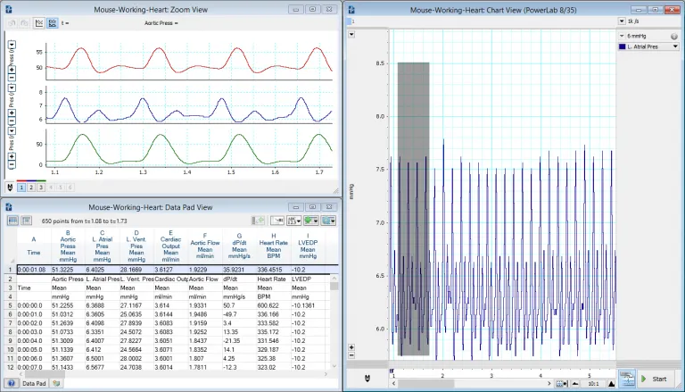
A typical Working Heart recording in LabChart (mouse model) featuring aortic, atrial and LV pressures, flow, and dp/dt.
Langendorff Perfusion
Unlike the working heart model, the Langendorff system relies on retrograde perfusion of the heart (through the coronary vasculature) in order to maintain cardiac function. This means the perfusate does not enter the left ventricle.
In brief, the technique involves cannulation of the aorta, which upon perfusion results in the closing of the aortic valve, thereby preventing flow into the left ventricle. Instead, the perfusate passes from the aortic root via the ostia into the coronary vasculature, providing the nutrients and oxygen required to maintain the physiological function of the heart. Typically, Langendorff perfusion is used to study ischemia-reperfusion injury, vascular reactivity, electrical conduction, and pharmacological interventions. Although the Langendorff preparation does not mimic real-life physiology as closely as a Working Heart model, it has the advantage of a simpler and more cost-effective preparation that can be suitable for many applications.
Langendorff available parameters include:
- Left ventricular developed pressure
- Perfusate pressure
- ECG/EKG
- Heart Rate
- Temperature
- Oxygen, carbon dioxide and pH
- Coronary flow
- Pacing
See here for more information about our Langendorff solutions.

A typical Langendorff recording in LabChart (rat model) featuring LVP, ECG, perfusion pressure and calculated variables.
Related: Effects of constant flow vs. constant pressure perfusion on Langendorff isolated heart studies.
Myocyte Isolation
For some researchers, the goal of an isolated heart study may not be to record data from the entire heart, but to isolate cardiomyocytes for further analysis. Cardiomyocytes are cardiac muscle cells that are responsible for the contractile forces of the heart. For myocyte isolation, a simple Langendorff perfusion can be used to remove the myocytes from the surrounding extracellular matrix. After being isolated, there is a vast amount of applications studied from pharmacology or toxicology, or specifically, cell mechanics, immunohistochemistry, calcium imaging, and many more. To isolate the cells, an intact heart must first be isolated and maintained through a perfusion system.
We offer various configurations based on your specific needs and goals, such as what enzyme/s you are using:
- Single or multiple reservoirs system
- Gravity fed or peristaltic pump driven
- Recirculating vs non-recirculating
- High Yield and Low Volume
- Heart or tissue bath chambers
Please contact your local ADInstrument's sales representative or office to talk to our team about myocyte isolated collection techniques, systems and equipment.
