
Get the right tool for your research measuring invasive blood pressure.
When collecting data from acute invasive pressure studies, it is essential to have the right set of tools at your disposal. Ensure that you know the differences between the available technologies to get the right equipment for your needs. You might be familiar with pressure catheters already. Solid-state pressure catheters are the gold standard for measuring real-time pressure. Fluid-filled (or gel-filled) catheters are a cost-effective and robust alternative. Is it time to upgrade from a fluid-filled to a solid-state pressure catheter?
In this article, we will cover the main differences:
- Specifications
- Data Accuracy
- Experimental applications
- Investment
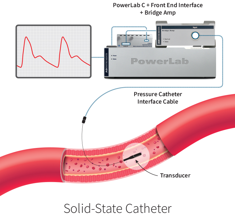
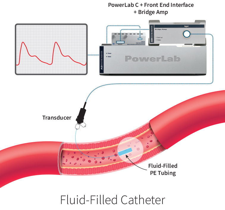
Figure 1: Illustration of the Solid-state Catheter and Fluid-filled Catheter pressure recording setup. The solid-state pressure transducer is within the catheter tip at the site of interest, and the pressure waveform shows a sharper systolic peak and a pronounced dicrotic notch. In contrast, the fluid-filled pressure transducer is outside the body.
Provide your details below to receive the full version of this article.
Specifications
Transduction Site

Solid-state pressure catheters contain a signal transducer and a MEMS sensor - a silicon chip that flexes with each pressure pulse - within the catheter tip at the site of interest.

Fluid-filled catheters translate each pressure pulse along the length of the fluid medium within the catheter to the pressure transducer located outside of the animal.
Size
The size and shape of your catheters are critical to experimental design, particularly considering the diameter of veins within animal models such as rats or mice.
The polyethylene (PE) tubing utilized determines the size of a fluid-filled catheter. Various dimensions are available (PE50 ~3 FR, PE10 ~2 FR). Smaller catheters in larger animal models may introduce artifacts in signal due to movement in the bloodstream.
Care
Cleaning and handling are essential parts of ensuring equipment fidelity.
Blood clotting can occur surrounding the solid-state sensor; however, signal attenuation is negligible. Proper device cleaning and careful handling and storage will ensure the device’s longevity.
Blood pressure transducers for fluid-filled catheters should be cleaned between uses. Fix any blood exchange into the catheter medium with a catheter flush to prevent clots from attenuating the pressure signal.
Reusability
Both fluid-filled and solid-state pressure catheters require calibration with a pressure gauge before each experiment.
Solid-state pressure catheters are reusable given proper care and cleaning, with no disposable elements.
Researchers should replace PE tubing for a fluid-filled catheter between each experiment. It is inexpensive and can be cut to length to match requirements. Blood pressure transducers are reusable when cleaned correctly.
Data Accuracy
Data accuracy is the primary motivator for researchers upgrading from a fluid-filled to a solid-state pressure catheter.
Solid-state pressure catheters give higher quality data for invasive blood pressure monitoring, measuring directly at the site of interest.
Fluid-filled catheters measure pressure indirectly, relying on a pressure wave signal transmission through the fluid medium to the transducer.
Attenuation
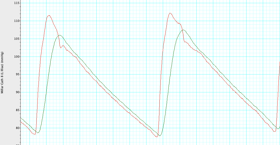
Figure 2: Waveform precision when utilizing a solid-state pressure sensor at the catheter tip, measured with LabChart8 software (1). A comparison of blood pressure waveforms recorded using fluid-filled catheters (green) and the Millar Acutip pressure catheter (red) showed attenuation of the blood pressure signal measured by the fluid-filled pressure catheter. In particular, there is a dampening of systolic amplitude and no pronounced dicrotic notch.
Solid-state pressure catheters record sharper peaks and a more pronounced dicrotic notch in cardiac measurements (Figure 2), data typically lost due to the significant signal attenuation of fluid-filled catheters. This resolution is critical for accurate systolic and diastolic pressure data.
Fluid-filled catheters are commonly relied on for Mean Arterial Pressure where signal attenuation has a limited effect. For experimental designs which analyze blood pressure waveforms, signal attenuation due to catheter diameter, high-frequency pressure pulses, or bubbles within the fluid may compromise data quality (2).
Artifacts
Noise and artifacts can severely distort blood pressure waveform and compromise data quality.
The pressure waveform recorded by solid-state pressure catheters is unaffected by motion, clotting, gravity, catheter flushing, or catheter bending - given proper cleaning.
Fluid-filled catheters may incorporate these artifacts into the measured pressure waveform, reducing the reliability of such data.
Phase Shift
Phase shift is detrimental to studies where precise, real-time physiological measurements are needed. This occurs when the pulsatile event and measurement of that event are desynchronized.
Data recording devices that transduce pressure signals directly at the site of interest, such as solid-state pressure catheters, are unaffected by phase shift.
Data recorded from a fluid-filled catheter can be affected by a phase shift. The length of the catheter tubing causes this phase shift, as the pressure signal takes time to reach the blood pressure transducer.
Frequency Response
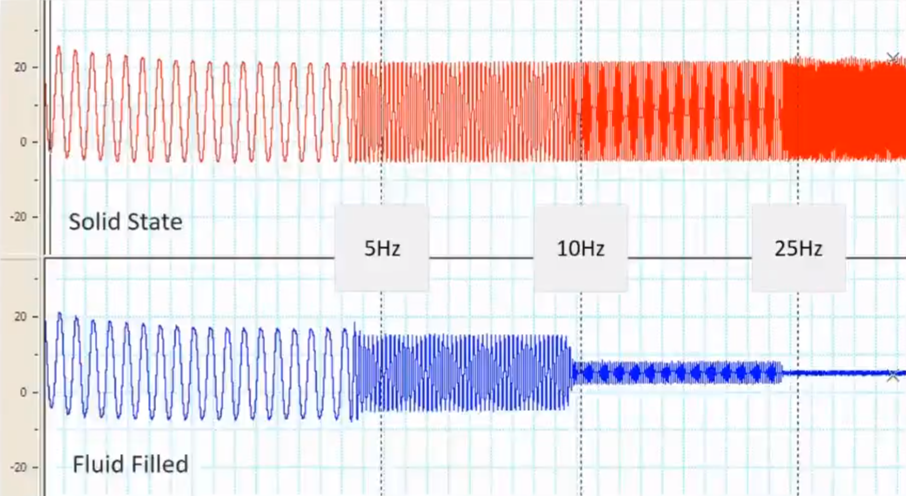
Figure 3: Maintain superior frequency response when utilizing a solid-state pressure catheter (3). A comparison of frequency response recorded using fluid-filled catheters (blue) and the Millar Mikro-Tip® pressure catheter (red) showed attenuation of the signal at higher frequencies when measuring a speaker-produced wave within a fluid-filled chamber.
Frequency response describes the range of frequencies over which the pressure waveform will experience minimal attenuation. In invasive blood pressure studies, the frequency response should be high enough to measure elevated heart rates in your animal model - particularly in rats or mice.
Solid-state pressure catheters can measure frequency into the kilohertz range without experiencing any signal attenuation.
Fluid-filled catheters can lose waveform amplitude at higher frequencies (Figure 3), affecting data quality.
Experimental Applications
If you are setting up a new protocol or are interested in upgrading your invasive pressure monitoring equipment, investing in equipment suitable for multiple applications is advantageous.
The sensitivity of solid-state pressure catheters enables measurement of intracranial (4), intravesical (bladder) (5), compartment, and tumor pressure.
Fluid-filled catheters generally do not have the sensitivity and signal fidelity for use in non-cardiac research.
Investment
Price is always a factor when choosing the right equipment for your research.
Solid-state pressure catheters are a premium product, with the data quality to match.
Fluid-filled catheters are cost-effective, robust, and widely available.
Conclusion
Upgrading to a solid-state pressure catheter from a fluid-filled catheter will vastly improve your data quality.
Solid-state pressure catheters are:
- Available in multiple sizes and shapes, suitable for various model animals.
- Reusable, given proper care and storage.
- Highly accurate, with reduced waveform attenuation for precise systolic and diastolic pressure measurements.
- Unaffected by artifacts and phase shift.
- Suitable for measuring frequencies in the kilohertz range, with high frequency response for maintaining waveform fidelity in small animal models.
- Used for applications beyond cardiac pressure monitoring, such as intracranial and intravesical pressure.
RECOMMENDATION:
We recommend Millar Mikro-Tip® Pressure Catheters for accuracy and reliability.
Fluid-filled catheters are robust devices that provide more insight into arterial blood pressure than non-invasive measures. However, if your research requires accurate pressure waveform analysis, solid-state pressure catheters such as the Millar Mikro-Tip® provide unbeatable signal fidelity.
Need more information? Listen to this webinar with Dr. Tom Smith, an expert in microsurgery and vascular pressure measurement techniques.
Need to record long-term invasive pressure measurements? Read “Solid-state sensors at the catheter tip – Why settle for less?” to discover the power of Millar Mikro-Tip® Pressure Catheters combined with wireless telemetry technology.
Provide your details below to receive the full version of this article.
Related products:
Contact us to discuss which pressure measurement device best suits your research and workflow.
Helpful Resources
- Figure from data collected by ADInstruments
- Vecchi, Adelaide de, et al. “Catheter-Induced Errors in Pressure Measurements in Vessels: An In-Vitro and Numerical Study.” IEEE Transactions on Biomedical Engineering, vol. 61, no. 6, 2014, pp. 1844–50. Crossref, https://doi.org/10.1109/tbme.2014.2308594.
- Slide from ADInstruments/InsideScientific webinar “Invasive Blood Pressure – fundamentals and best practices for preclinical research” presented by Dr. Tom Smith.
- Thakkar, Pratik, et al. “Hypertensive Response to Ischemic Stroke in the Normotensive Wistar Rat.” Stroke, vol. 50, no. 9, 2019, pp. 2522–30. Crossref, https://doi.org/10.1161/strokeaha.119.026459.
- Hunter, Diana V., et al. “Preserved Adrenal Function After Lumbar Spinal Cord Transection Augments Low Pressure Bladder Activity in the Rat.” Frontiers in Physiology, vol. 9, 2018. Crossref, https://doi.org/10.3389/fphys.2018.01239.