How the dead teach the living
Donating your body to science could be the last thing you do on this earth - a selfless act that could change the lives of many.
While many medical educators still consider cadaver labs as the gold standard for teaching anatomy, rapid advancements in technology over the last 20 years has led to the development of virtual dissections - a popular alternative to the traditional ‘hands-on’ method.
Replacing the scalpel with a computer mouse, students are able to view anatomical structures from any angle, zoom in on hidden structures, and simply click ‘reset’ if they make a mistake. To understand how these digital cadavers are made and their impact on medical education - we talked to Rachel Klaus from Touch of Life Technologies (ToLTech), a company that creates and builds 3D models from thousands of cross-sectional images of human bodies - all in the name of science.
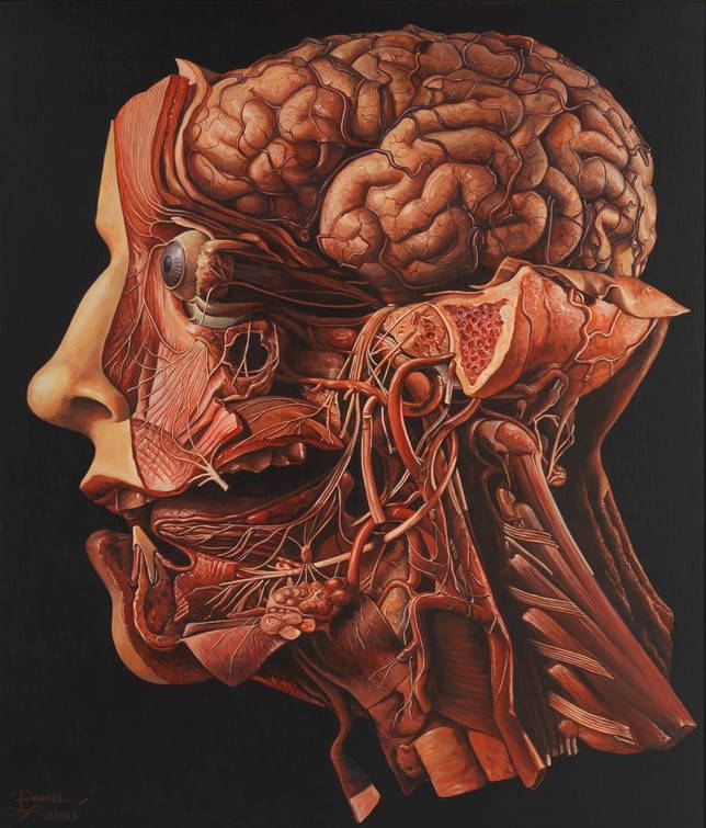
A human head dissected. Credit: Wellcome Collection. CC BY.
The Visible Human Project
The original Visible Human Project was initiated in 1991 when the contract to perform the work was awarded to Drs. Spitzer and Whitlock at the University of Colorado School of Medicine. In 1993 the first body was sectioned and imaged from head to toe. Funded by the National Library of Medicine, the vision of The Visible Human Project was to create a highly detailed image database of male and female human anatomy to be used as a foundational model for education and medical imaging research.
The first body to be sectioned as part of the project was that of a male prisoner from Texas who was executed. Shortly after his death, his body was scanned from head to toe using clinical MRI and CT imaging before being encased in blue gelatin and frozen to preserve the tissue. To slice and image the cadaver, they had to section the body into 4 blocks to fit within the cryomacrotome (milling machine). Here, sections of each block could be removed 1mm at a time and a high-resolution color picture taken of each newly exposed surface. By taking such small sections, and aligning the thousands of pictures taken, an extremely detailed map of the human body was created, uncovering the intricate network of nerves and blood vessels surrounding the organs and muscles of the body at an astounding level of detail.
‘ToLTech was founded to build education and training tools based on real anatomy from The Visible Human Project and other high-resolution cross-sectional datasets. We developed the VH Dissector software, that allows you to navigate the original cross-sectional views of the body and also create views of the anatomy in other cross-sectional planes and in 3D. This is unique in comparison to other software programs as everything visualized is real, photographic anatomy from an actual human cadaver as opposed to artistic renderings and models created by illustrators. This provides a sense of reality not found in other platforms.’
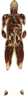
Susan Potter
ToLTech’s VH Dissector software allows you to rotate and visualise the human body from any angle through 3D and cross-sectional views of real anatomy.
An elderly woman from Denver, Colorado read about The Visible Human Project in the newspaper and decided she wanted to donate her body for similar educational purposes. Susan Potter wasn’t a healthy individual by any means. She had undergone multiple surgeries and been afflicted with numerous health issues such as breast cancer, resulting in a double mastectomy and a hip replacement. Despite this, it was her wish to dedicate her body to enhance the learning of future medical students and further our understanding of the human body.
‘Vic initially said to Susan “no I'm not going to do this”, but she was persistent enough that he eventually agreed. She promised to provide documentation of her life in addition to her body after death. She also assured Vic that she would die within a year, but then she lived for 12. She passed away in 2015. I never knew her while she was living.’ It wasn’t until two years after her death that Susan began her journey to become a visible human.
For more information, check out the National Geographic video and article following Susan’s journey.
Susan was sectioned into more than 27,000 slices, 1/16 of a mm thick, over 60 days - generating over 2 terabytes of images. ‘When we cut it's a 24/7 process - It takes a lot of coordination and dedication. We like to get students who express interest involved as well.’
Check out the short video below of the cryomacrotome in action!
How do you create a 3D model from thousands of individual 2D images?
‘We have developed our own software that allows us to see the cross-sections in the three planes; sagittal, coronal (or frontal) and transverse. Then we can go through and lay down 3D points to define each anatomical structure. So we essentially trace an artery, vein, or other structure by overlaying these points on the cross-sectional pictures to create a 3D model.’
Related: 10 free high-quality anatomy images to download and use »
Who identifies the different parts of the body? A person or a machine?
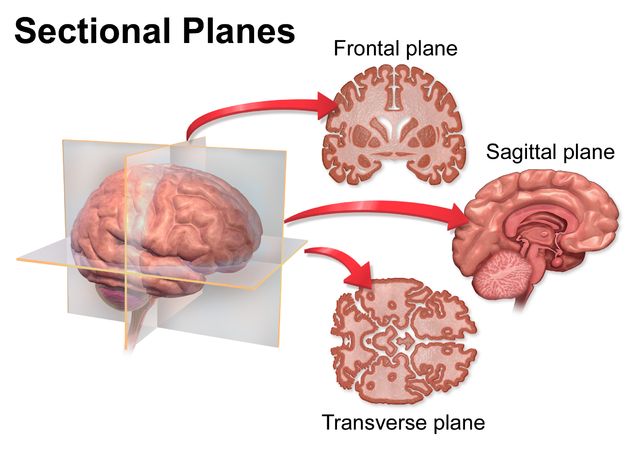
Anatomical Axes. Credit: Blausen.com staff (2014).
‘Identification of high contrast structures (e.g. bones) is done by machine, but all the detail work is done by people. We hand identify, outline and label each cross-section. That’s where technology still hasn't caught up with what we are trying to do. We can't really automate much of it, as it's all too variable and requires precise identification to accurately label each structure. This process is different to how clinical images are automatically segmented or visualized.’
So how long does it take?
‘It takes a lot of manpower. We are still working on the original two visible humans, updating the models with improved technology. We haven't released any of Susan Potter’s data because there's a lot that goes into making something usable. You have to assemble and edit the >27,000 images, identify anatomical structures and all their boundaries in order to make the models. Then you have to check the models and their relationships to other models and ensure their display works the way it is supposed to.’
‘It is really difficult to find people who can do this kind of detailed modeling work. We have students and full-time employees working on the data, but we can always use more. It's one of those things where “the scientist is never done with their work”, there's always more to fix and perfect. It’s a never-ending project. By the time we get this perfected, there will probably be a new way to model - hopefully, it’ll be automatic…!’
Do you think digital cadavers will replace traditional dissections?
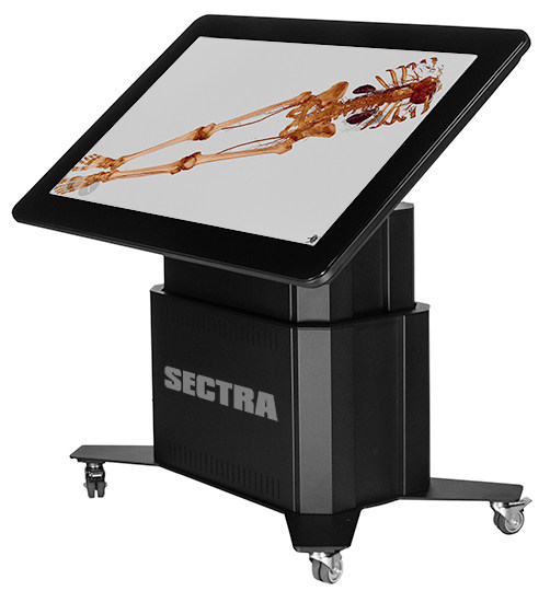
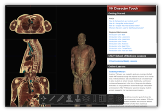
The Sectra virtual dissection table, developed in conjunction with ToLTech.
‘That is a hot topic right now. I think that eventually it will, but whether that's in 50, 100 or 1000 years I have no idea. There are a lot of people who think dissecting is the gold standard for learning Anatomy, so we develop our software not with the intention to replace but to augment dissection. There are many things you can do in a virtual environment that cannot be done in a traditional lab and vice versa.’
‘The great thing about our software is that the model is always there and you can always click reset. When you’re dissecting a cadaver, one wrong cut and you will destroy what you were looking for, or damage the bit of tissue you were wanting to look at. However, in saying that, our software does not allow interaction with tissue the same way a cadaver does. It’s digital. If you want to remove the muscle, you have to remove the entire muscle, you can’t peel it back.'
'There are obviously limitations to the technology, which is one reason it hasn’t replaced classic dissection, but who knows what technology will do in another 100 years. In saying this, a few schools are already using our products to replace dissection - It’s only a matter of time before more and more start trending this way.'
Are you looking for high quality dissection imagery and videos for your anatomy course?
Created in partnership with ToLTech, the Lt Anatomy Collection is a complete curriculum of ready-to-use lessons and labs featuring media-rich dissection and histology images and videos from teaching and assessments. Combine with our Physiology Collection, and you have a complete A&P course on a single delivery platform.
Click here to take a look inside the Anatomy Collection »
Related: Check out some of the top features from our favorite dissection and histology labs, the Heart Dissection lab and the Bone and Cartilage Histology lab.
More for anatomy educators
Anatomy Month: Anatomy and Art: Leonardo da Vinci – The science behind the paintings
Resources for Educators: The HAPS Anatomy Learning Outcomes
Free Images: 10 free high-quality anatomy images for educators to download and use!
Anatomia Italiana: Professional Development in Italy that combines anatomy and art